

Why flexural waves are the future of biomechanics
Using an elegant law of mechanics, engineers have discovered how to measure the compressive force on the tibia. Their wearable device could revolutionise biomechanics

Every stride a runner takes sends a shockwave of force rippling up through the skeleton. In the tibia — the main load-bearing bone of the lower leg — that force can reach fourteen times bodyweight during a sprint.
Athletes, clinicians and researchers have long wanted to track that load in real time, to catch the early warning signs of stress fractures, to optimise training programmes and to understand the precise mechanics of human gait. But actually measuring the force inside a living bone, while that person is out on the road rather than wired up in a laboratory, is difficult.
The best available wearables — accelerometers strapped to the shin, pressure insoles in the shoe — capture only rough proxies for bone loading, and decades of research have shown these proxies can be unreliable.
What researchers would dearly love is a non-invasive way to make real world measurements much more accurate.
Compressed beam
Now Ali Yawar and colleagues at Harvard University in Cambridge, have found an elegant solution hidden in an old branch of mechanics: the way sound propagates through a compressed beam changes in a predictable, measurable way as the compression force varies. The human tibia, it turns out, behaves much like such a beam.
They demonstrate that the tibia behaves like a compressed beam whose vibrational properties change depending on the weight it supports. By mounting a small mechanical “tapper” and an accelerometer to the skin, the team can measure how “flexural waves” travel through the bone. As the compressive load on the tibia increases, the frequency spectrum of these waves shifts in a predictable, linear fashion. “Tibial compressive loading, like loading in other bones, impacts maintenance, growth, and injury, and is therefore a key variable to measure in experimental human biomechanics,” they say.
The approach is rooted in Euler-Bernoulli beam theory, which describes how slender, elastic beams respond to bending loads. When a compressive force is applied along the beam’s axis, it selectively stiffens the beam against low-frequency bending more than against high-frequency bending. This shifts the spectrum of flexural waves travelling along the beam towards higher frequencies as the compressive load increases, and it does so linearly for the physiological range of forces seen in the human tibia.
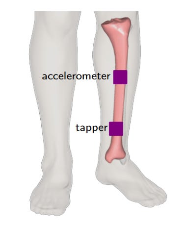
The Harvard team built a wearable system to test this effect. A small bone-conduction audio transducer, strapped to the leg near the ankle, taps the tibial shaft fifty times per second, each tap launching a brief flexural wave along the bone.
A piezoelectric accelerometer mounted 16 centimetres up the shin then measures each wave as it travels up the bone, revealing how the frequency of the spectral peak shifts as the tibial load waxes and wanes. By tracking that peak in real time, the system tracks the compressive force.
The team tested the device on five men and four women as they performed two tasks on an instrumented treadmill: slow lateral swaying, which loads and unloads the tibia rhythmically at about 0.2 Hz, and walking at 0.4 metres per second.
They then compared the results with calculations from computer model of human motion fed with optical motion capture and force-plate data.
The agreement was striking. “Data from nine participants demonstrate linear relationships between tibial compressive force and spectral peak location,” say Yawar and co, exactly in agreement with beam theory.
The authors are candid about current limitations. For more widespread use, the device will need to be shrunk and optimised. They will also need to develop a simple way of calibrating the device at the beginning of each measurement session. And the sensor cannot be used on body sites where thick soft tissue attenuates the signal, which might limit its use for some individuals.
Nevertheless, the study establishes proof of concept for a fundamentally new way of sensing. “This flexural wave-based technique could give rise to a new class of wearable sensors for non-invasive physiological bone load monitoring and measurement, impacting research in human locomotion and sports medicine,” say Yawar and co. “The non-invasive and portable nature of our technique has implications for the development of new wearable sensors for in-field measurements and continuous monitoring of bone loading in athletic, clinical, or industrial environments.”
Ref: arxiv.org/abs/2511.06140: Non-invasive load measurement in the human tibia via spectral analysis of flexural waves
INSIGHT
This paper introduces a fundamentally new approach to measuring internal bone loading, with implications that extend well beyond tibial biomechanics. Its new insight — that bones can effectively be “listened to” for real-time load information — opens a new measurement paradigm applicable in principle to any long bone in the body.
This makes wearable bone-load monitoring possible in almost any setting. Athletes could receive instantaneous alerts when tibial loads exceed safety thresholds, potentially preventing stress fractures before they occur. Industrial workers could be warned before repetitive strain injuries take hold.
The approach also has the potential to be adapted to other long bones, such as the femur or humerus, creating a comprehensive map of skeletal stress during complex movement.
Beyond load, the frequency response of flexural waves is governed by Young’s modulus and bone geometry and other researchers have exploited this to measure bone composition. The new tapping technique hints at the possibility of using wearable sensors to monitor changes in bone mineral density or healing progress in fractures over time.
And in better understanding body loads, the technique could help improve next generation of intuitive prosthetic limbs and industrial exoskeletons.